

Today, December 20, 2013, I had my first appointment with the pain management doctor.
I’m happy to report that it was a good experience. I was apprehensive because I was meeting with a new health provider. I knew that this was my last option because I have already tried different types of therapy, including alternative, to obtain relief from the back pain that started in May 2013.

I had an MRI done which was reviewed with my primary care provider.
~~Impression~~
1. Small, left sided posterolateral disc protrusion at L5-S1 causing some narrowing of both neural foramina
2. Bulging disc at L4-5, L3-4, L1-2 and T12-L1
3. Dextroscoliosis with global disc dessication (drying up, loss of hydration), degenerative end plate changes
Needless to say, this looked ominous! And, I was expecting the worse.

The doctor turned out to be very professional and, most of all very well prepared and caring. He asked the pertinent questions and performed a physical examination.
He explained everything in detail and listed options. We are going to follow logical steps. We will start with the basics. The changes found in my vertebral spine are not going to change. But symptoms can be treated conservatively.
Treatment is aimed to reduce inflammation of surrounding tissue, improve quality of life and increase mobility, hence, physical activity.
In spite of the description of the MRI results, it wasn’t as bad as it seemed.
The pain is localized and is not associated with neurological symptoms.
This is an excellent sign.

A Patient’s Guide to Lumbar Facet Joint Arthritis

Introduction
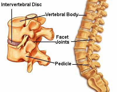
Arthritis of the lumbar facet joints can be a source of significant low back pain. Aligned on the back of the spinal column, the facet joints link each vertebra together. Articular cartilage covers the surfaces where these joints meet. Like other joints in the body that are covered with articular cartilage, the lumbar facet joints can be affected by arthritis.
The human spine is made up of 24 spinal bones, called vertebrae. Vertebrae are stacked on top of one another to create the spinal column. The spinal column gives the body its form. It is the body’s main upright support.
The back portion of the spinal column forms a bony ring. When the vertebrae are stacked on top of each other, these bony rings create a hollow tube. This tube, called the spinal canal, surrounds the spinal cord as it passes through the spine. Just as the skull protects the brain, the bones of the spinal column protect the spinal cord.
Between the vertebrae of each spinal segment are two facet joints. The facet joints are located on the back of the spinal column. There are two facet joints between each pair of vertebrae, one on each side of the spine. A facet joint is made of small, bony knobs that line up along the back of the spine. Where these knobs meet, they form a joint that connects the two vertebrae. The alignment of the facet joints of the lumbar spine allows freedom of movement as you bend forward and back.
The surfaces of the facet joints are covered by articular cartilage. Articular cartilage is a smooth, rubbery material that covers the ends of most joints. It allows the bone ends to move against each other smoothly, without friction.
Related Document: A Patient’s Guide to Lumbar Spine Anatomy
Causes
Why do I have this problem?
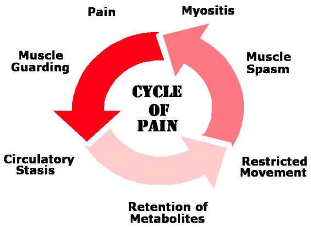
Normally, the facet joints fit together snugly and glide smoothly, without pressure. If pressure builds where the joint meets, the cartilage on the joint surfaces wears off, or erodes.
Each segment in the spine has three main points of movement, the intervertebral disc and the two facet joints. Injury or problems in any one of these structures affects the other two. As a disc thins with aging and from daily wear and tear, the space between two spinal vertebrae shrinks. This causes the facet joints to press together.
Facet joints can also become arthritic due to a back injury earlier in life. Fractures, torn ligaments, and disc problems can all cause abnormal movement and alignment, putting extra stress on the surfaces of the facet joints.
The body responds to this extra pressure by developing bone spurs. As the spurs form around the edges of the facet joints, the joints become enlarged. This is called hypertrophy. Eventually, the joint surfaces become arthritic. When the articular cartilage degenerates, or wears away, the bone underneath is uncovered and rubs against bone. The joint becomes inflamed, swollen, and painful.

Full article/Source: http://www.methodistorthopedics.com/lumbar-facet-joint-arthritis
After the doctor’s full explanation, I asked: “When can we do this?”
He said …. “Right now”. Let’s get this ball rolling!!
Initially I had an IV inserted, received conscious sedation (a medication called Versed) and was taken to the procedure room.
Today I underwent injection of four facet joints at the level of the lumbar spine, two on each side. The procedure was done under fluoroscopic control which means that the doctor used an Xray machine to know exactly where he needed to inject the medication.
What is a lumbar facet joint block?
A lumbar facet joint block is an injection of local anesthetic (numbing medicine) into one or more of the small joints located along the side of each vertebrae on both sides of the spine in the lower part of the back. Multiple injections may be performed, depending upon how many joints are involved.
Facet joint blocks are typically requested for patients who have pain primarily in their back as a result of arthritic changes in the facet joints or for patients who have mechanical low back pain. A facet joint block may be diagnostic (a test to see if your pain is coming from this area) and/or therapeutic (to relieve your pain).

How do I prepare for the procedure?
No solid food or fluids after midnight prior to the procedure unless directed otherwise. You may take your medications with a small amount of water. Diabetics should not take their medication for diabetes until after the procedure is complete. Please check your blood sugar at home before arriving at the PMC. If you are taking any blood thinners such as Coumadin, Warfarin, Plavix, or any others, these medications must be discontinued well before the procedure.
You will be directed by our staff as to when you should stop this medication. Please make your Pain Management doctor aware that you are taking a blood thinner, and contact your primary care physician or prescribing physician before stopping this medication.
What are the risks of the procedure?
As with most procedures, there is a remote risk of bleeding, infection, nerve injury, or allergic reaction to the medications used. Other short-term side effects may occur. If local anesthetic spreads to nearby nerves you may have weakness or numbness that can last for several hours. If this happens, you may have to stay in the Pain Management Center until it resolves.
You may have increased pain for a few days after the injections, including localized pain at the injection site.
What happens during the actual procedure?
The procedure will be done in the fluoroscopy (X-ray) room with you lying on your stomach. An intravenous (IV) line may be started in your hand or arm to give fluids and/or medication to help you to relax. Your back will be thoroughly cleansed with an antiseptic soap. Sterile drapes will be placed over your lower back. Your back will be numbed with injections of local anesthetic using a very small needle. You may feel a brief stinging or burning sensation, which will go away in about 15 seconds. Using X-ray guidance, longer needles are then advanced into the facet joints along the spine. Once the needles are in the proper location, a local anesthetic medication (with or without steroids) will be injected, and the needles will be removed. Your skin will be cleaned again, and Band-Aids will be applied.
You will be moved to the recovery area, where your vital signs will be monitored for an appropriate time, usually about 20-30 minutes. You will be given verbal and written discharge instructions, and you will be able to leave with your driver after your doctor authorizes discharge.
How will I feel after the injection?
Your back pain may be improved immediately after the injection as a result of the local anesthetic. It is important to keep track of how you feel for the rest of the day. We encourage you to move around and do your usual activities, provided they are not too strenuous.
It is important that you keep track of the amount of pain relief you receive as well as how long the pain relief lasts.
Some local tenderness may be experienced for a couple of days after the injection.
Using an ice pack three or four times a day may help alleviate this. We would like you to hold off taking pain medication the day of procedure so you will be able to accurately see how much of your pain is relieved by the procedure alone. Your feedback about pain relief after the procedure will guide us in deciding the next step in your treatment.
Will I have any restrictions on the day of the procedure?
You may not drive for the remainder of the day after your procedure. A responsible adult (over 18 years old) must be present to drive you home or to go with you in a taxi.
The procedure will be cancelled if you don’t have a responsible adult with you! This is for your safety.
No heat is to be used on the injected area for the remainder of the day. No tub bath, shower or soaking in water (i.e., pool, hot tub, etc.) for the remainder of the day. You may resume normal diet and medications after the procedure unless told otherwise by your doctor.
Read more: http://www.kernan.org/pain/lumbar_facet_joint_block_faq.htm#ixzz2o4MV6s1W
~~The next option it facet intra-articular injection isn’t effective~~
Facet Rhizotomy
Rhizotomy describes a surgical procedure in which a nerve is purposely cut or destroyed. Facet rhizotomy involves severing one of the small nerves that goes to the facet joint. The intent of the procedure is to stop the transmission of pain impulses along this nerve. The nerve is identified using a diagnostic injection.
Then the surgeon inserts a large, hollow needle through the tissues in the low back. A special probe is inserted through the needle, and a fluoroscope is used to guide the probe toward the nerve. The probe is slowly heated until the nerve is severed.

Source: http://www.spinesurgeon.co.uk/content/radioneurot/
Let’s not get ahead of ourselves yet.
For now I’m doing better. I am still sore but that is a natural effect. The test will be tomorrow when I have to get out of bed in the morning.
Facet Joint Steroid Injection
In the meantime, if this is effective, the quality of life will improve. I will be able to be more active and have no pain or decreased intensity in the pain. I’m hopeful … This will get better. It has to ….
I WANT THE PAIN TO GO AWAY!! I WANT TO BE NORMAL AGAIN!!

We ALL are ONE!!

